SEM & EDS Services
for Quality
Analysis and to Resolve Quality Issues
Whether it is a quality or reliability problem, or a research and development question, Houston Electron Microscopy delivers answers understandable to any audience. Houston Electron Microscopy, Inc. specializes in SEM Microscopy and EDX Spectrometry, and is dedicated in providing you with the technical data and information that will lead to answers.
We can help quality control engineers and scientist with product contaminant, surface defects, and coatings issues. We can identify the contaminant and the size and shape of the contaminant. Coating and coating substrates can be measured and examined down to the micron level.
A Scanning Electron Microscope, (SEM) exposes microscopic properties of particles, including chemical composition, morphology, and contaminant identification. We can observe microscopic features in any solid material at extremely high magnifications. Particles and features in the nanometer range can be observed and photographed.
With the integrated Energy Dispersive Spectrometer (EDS) system allows us to analyze the elemental composition of any feature observed under the SEM. Oils and liquids can be filtered, dried and analyzed for particle identification and chemical composition. Elemental and compound information can be reported in several formats:
•Qualitative elemental spectrums showing the elements present
•Quantitatively showing the atomic and weight percent
•X-Ray mapping showing the concentration
and location of the elements present overlaid onto the SEM image
Our scanning electron microscopy laboratory has the necessary equipment for the preparation of specimens for SEM/EDS analysis; from sawing to metallography to gold sputtering. Same day or next day service is available in most cases.
Examples of Quality Applications

Failure Analysis
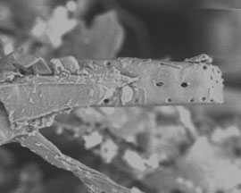
SEM examination of the fractures, wear surfaces, and corrosion deposits can lead to the cause of failure of many different metals.

Powders and Particle Analysis
Fracture surfaces of hard materials such as diamond aggregates (PDC) and WC can be examined for leach depth and phase distribution.

Coatings Analysis
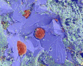
With the Scanning Electron Microscope, coatings of any type can be examined to the finest detail for composition and morphology, thickness, porosity, layers, pinholes, etc.

Powders and Particle Analysis
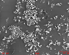
Image of a diatom among dust particles. Our Scanning Electron Microscope system is capable of imaging at high magnification with a deep field of focus.
Experience in Failure Analysis and Materials
Our primary goal is to get you the answers to your materials problems.
Example Applications
- Failure Analysis
- Fracture surface characterization
- Corrosion deposit analysis
- Geological specimen and core analysis
- Coatings analysis and characterization
- Phase and particle analysis
- Polycrystalline Diamond Compact (PDC) analysis
- Particle contamination in oils
- Identification of process abnormalities
- Contaminant investigations
- Microstructural defect analysis
- Reverse engineering
- Welding engineering analysis
OTHER SERVICES
- Metallography
- Micro-hardness Testing
- X-Ray Diffraction
- FTIR analysis of organic materials
- Experience
- Modern SEM/EDS System
- No hidden charges
- Easy and quick availability
- Conveniently located in Northwest Houston
Advantages
We get answers
Houston Electron Microscopy
281-888-4261 or 281-704-0188 Fax:1.888.600.0212
Toll Free: 1-844-318-8775

Close up of
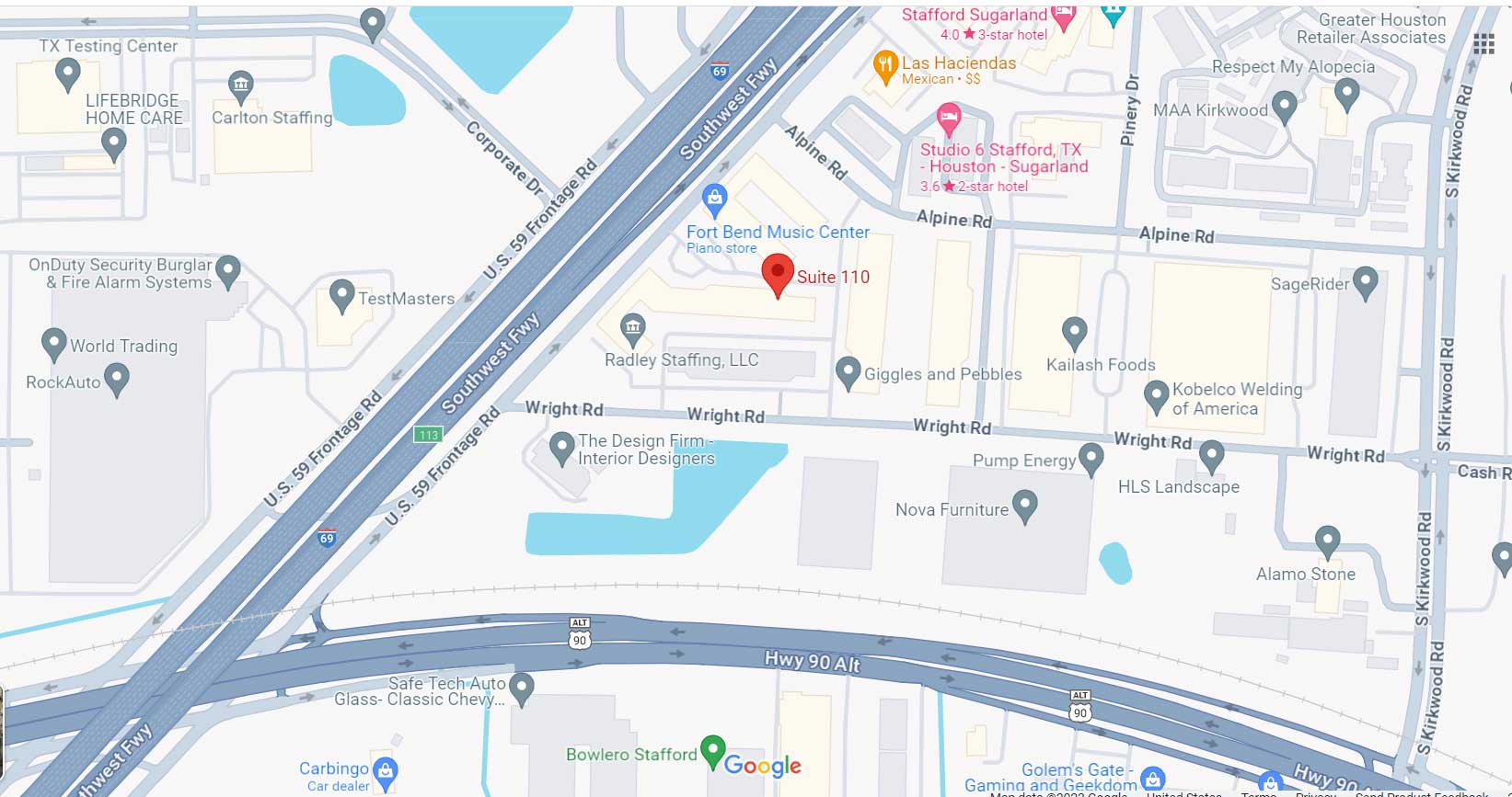
Our Location, (PDF file)


We get answers
281.888.4261